Correlation between organ dysfunctions and vertebral displacements
In a prospective study a comparison was made between a control group (a group of patients with displaced vertebrae but without organic complaints) and different groups of patients who were not only characterised by displaced vertebrae but also by various organ dysfunctions. Six groups of patients were differentiated: patients with gastric complaints, intestinal complaints, heart conditions, asthma, hyperventilation and migraine. Of these seven groups curves have been made to show the mean number of abnormalities per vertebra. The curves of patients with an organic complaint were compared with the curves of patients without organic complaints. The differences, which are pronounced, are discussed in this article.
Introduction
During the years that osteopathic medicine has been practised with an increasing degree of understanding and precision (Sickesz and Bongartz, 1989) there seemed to be a connection between positional deviations in the vertebrae and dysfunctioning in the dermatome, viscerotome or myotome.
Reasons for the study
Three groups of factors induced me at the end of the 1980s to set up a study into the connection between vertebral positions and organic complaints. These factors can be summarised as follows:
-
Practical experience
Prior to the treatment of every patient I made a note of all their complaints, not only complaints of pain but organ dysfunctions as well. After the treatment the complaints were reviewed with the patient. Frequently it was found that not only had the pain been relieved but also the dysfunctioning.
-
Publications
Publications can be found in the literature which point in the same direction. Kunert (1978) had already indicated a possible aetiological connection between vertebral column problems and organic dysfunction.
In the Netherlands, Van Cranenburgh (1987) has published an excellent book on this subject, while others have likewise devoted attention to this phenomenon (Ditmar and Dobner, 1961).
-
Anatomical-physiological considerations
Misaligned vertebrae are surrounded by oedema which is even perceptible in the sulcus paravertebralis at the point of the processus spinosus.
Certain vertebral abnormalities are accompanied by subluxations of the corresponding rib(s) in the costotransverse joint in the dorsocranial or dorsocaudal direction. This joint is traversed by the sympathetic trunk.
Sympathetic and parasympathetic efferent and afferent fibres are located within each intervertebral foramen, thus making a connection between vertebral abnormalities and vegetative stimulation probable.
Design of the study
In order to investigate more fully the suspicion of a connection between vertebral abnormalities and dysfunctioning in the dermatome, viscerotome or myotome, the decision was taken to conduct a prospective study.
-
Problem definition
Within the scope of this study it was investigated whether there were any differences in the number of misaligned vertebrae in patients without organic complaints and the number in patients with organic complaints. Specifically, for each vertebra of each patient the number of abnormalities was established and the data were presented in such a manner that a comparison of this number of abnormalities per vertebra in patients without organic complaints (control group) could immediately be compared with the number of abnormalities in patients who did have such complaints.
-
Which patients and which organic complaints
All new patients (i.e. patients who had never been seen by us before) were included in the study. As always, the first step was to make a note of all the complaints while the patient was still dressed; then the patient’s back was examined. Two doctors made the diagnosis of the back disorders. The manner in which this was done has already been published (Sickesz and Bongartz, 1989).
In view of past experience, the study was limited to a total of seven groups of patients: four groups who apart from neck and/or back complaints also had manifest organic complaints; a migraine group and a hyperventilation group were also involved in the study; lastly, a group of patients with only neck and/or back complaints was included. The total number of patients was 925. A number of patients had multiple complaints: intestinal complaints, heart disorders and gastric complaints, in particular, quite frequently occurred together. The patients (men and women of all ages) were included in the study in the order in which they presented at the surgery.
Breakdown of patients by complaint:
- 150 gastric patients. This group comprised patients with pyrosis, nausea, vomiting and recurrent ulcers and gastritis (curve 1).
- 163 asthma patients, all with the usual medication (prescribed by internists) (curve 2).
- 150 intestinal patients, ranging from frequent diarrhoea or distressing constipation to Morbus Crohn and ulcerative colitis (curve 3).
- 151 heart patients, ranging from arrhythmia to anginal complaints (curve 4).
- 133 hyperventilation patients. The frequency of the attacks varied from “occasionally” to “all day long” (curve 5).
- 150 migraine patients. The diagnosis was made in the medical circuit and the patients had the usual medication. The frequency varied from three times a week to three times a year (curve 6).
- 150 patients without organic complaints. This is represented by the lower curve in the graphs.
All patients were also suffering from neuralgic complaints, which was their reason for coming to the surgery.
A connection between vertebral problems and loss of renal function, menstrual disorders and fertility problems was not charted in a curve owing to an insufficient number of patients presenting with these disorders, although the results of the treatment certainly justify a similar study.
-
Method of data representation
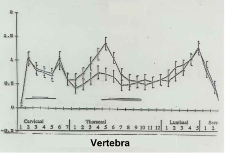
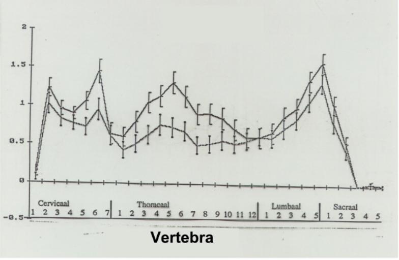
The data are presented with the aid of six graphs, one for each of the organic complaints. On the horizontal axis of each graph the vertebrae (C1 to S5) are indicated. The vertical axis shows the average number of deviating positions for each vertebra observed within the group of patients in question.
The number of abnormalities per vertebra is in the range of 0 to 3, with a sporadic peak of 4 abnormalities per vertebra. Where there were no abnormalities in the segment there was in very many cases a pelvic obliquity with a formula. Formulae are physiological adjustments which can degenerate into abnormalities if they become fixed owing to the pelvic obliquity becoming permanent (Sickesz and Bongartz, 1989).
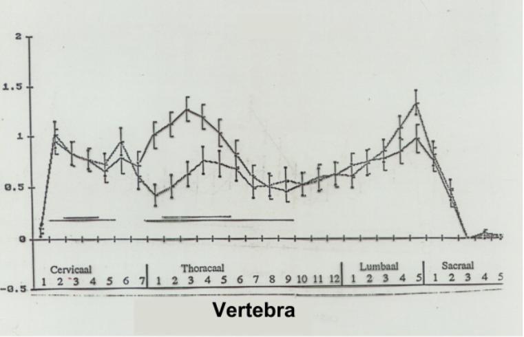
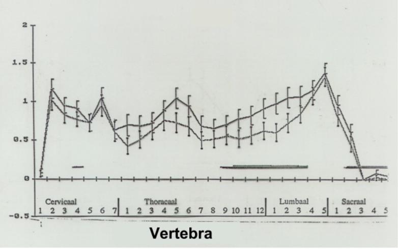
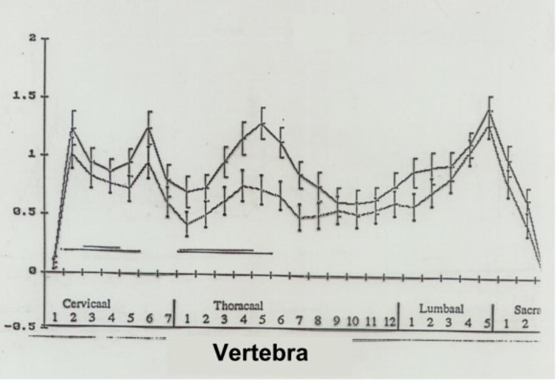
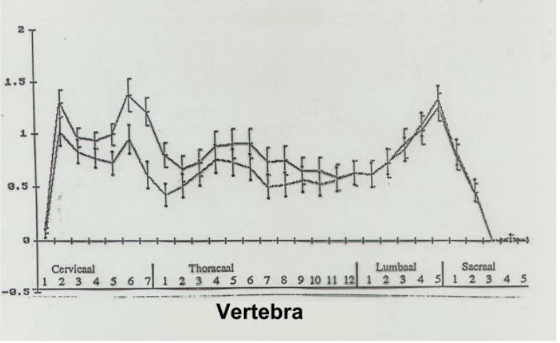
To simplify the comparison, in each of the six graphs the corresponding graph of the patients without organic complaints is included. The square brackets on the curves indicate the range.
Results of the comparisons
The lower curve in the graphs is the curve of the patients without organic complaints. All the graphs clearly show that than this curve is considerably lower across the whole spectrum than the curve of the patients with organic complaints. These patients therefore had fewer abnormalities in all segments. From this it may be concluded that in these patients the autonomic nervous system was stimulated less across the whole spectrum.
Diagram VII shows the relationship between the segmental efferent nerves and the internal organs as is known from anatomy. The segments that serve the organ in question are indicated in the curves with a line. The curves can now be divided into two groups, namely: curves 1 to 4, where the anatomical relationship between organ and segment is known, and the two dysfunctions hyperventilation and migraine, for which such a relationship is not known.
Curve I. Patients with gastric complaintsRadicular sensory impulses are conveyed to the stomach from segments T5 to T9 (Bumke and Forster). The curve shows that segments T3, T4, T5 and T6 have significantly more abnormalities in patients with gastric complaints than in patients without gastric complaints. The neck segments, however, do not show more abnormalities than in the control group. T5 is especially noticeable. In our experience, this vertebra is connected with the gastric juice content. Further research focused on changes in the gastric juice content before and after treatment is in preparation. |
|
Curve II. Asthma patientsThe radicular impulses to the lungs are conveyed from segments T1 to T5, with influences from T5 to T9. The curves show significant differences in segments T1 to T5. In practise, asthma in young children is found to respond well to vertebral correction. The paediatricians concerned have been contacted with a view to organising a study on a larger scale. |
|
Curve III. Patients with intestinal complaintsAccording to anatomy, the radicular impulses to the intestines are conveyed from T9 to L3. In segments T9 to L2 there is a significant difference between the two curves. A minor difference was also found to exist at T5. |
|
Curve IV. Patients with heart disordersThe radicular impulses to the heart are conveyed from segments T1 to T5 and segments C3 and C4 (n. phrenicus). Our curves show a significant difference at C6 and at segments T1 to T7. It may be said that the curves of the heart and the stomach resemble each other. That is in fact the case: the enervating segments actually overlap one other. The high peak at T5 is conspicuous. This vertebra was found to be therapeutically important in arrhythmia. In this case too, a study involving a large patient population should be conducted with ECGs before and after treatment. This publication may generate interest for such a study. Whereas the outcome of the curves in graphs 1 to 4 was supported by known data from anatomy, in the next two curves this is not the case since the segmental involvement in these disorders is not known. Perhaps these curves will prompt further investigation into this phenomenon. |
|
Whereas the outcome of the curves in graphs 1 to 4 was supported by known data from anatomy, in the next two curves this is not the case since the segmental involvement in these disorders is not known. Perhaps these curves will prompt further investigation into this phenomenon.
Curve V. HyperventilationIn this curve the vertebral abnormalities in segments T2 to T9 are seen to differ significantly from the vertebral abnormalities in patients without hyperventilation. Hyperventilation is the result of misalignments in the position of vertebrae and ribs, causing overstimulation of the autonomic nervous system. The complaint usually disappeared after correction. |
|
Curve VI. Migraine patientsA significant difference was found to exist in patients with migraine in respect of abnormalities in segments C5, C6, C7 and T1. The peak is situated at C7 and T1. One can imagine that the ganglion stellatum on the head of the 1st rib plays a role here. From the foregoing it may be concluded that the positional abnormalities of C7 and T1 play a role in the onset of migraine. The results of treatment are encouraging (Albers and Keizer, 1990). In this case too, a larger scale investigation should be organised. Apart from the correlation found here, no other objective abnormalities are known in migraine. The success of the therapy can therefore only be measured by subjective data from the patient with regard to the non-materialisation of attacks (Albers and Keizer, 1990). With a grant from the Ministry of Welfare, Health and Cultural Affairs a study has been conducted in recent years into the results obtained with osteopathic medicine. Many colleagues contributed data for this study. On 14 December 1990, E.D. Keizer and W. Albers obtained their doctorate at the University of Rotterdam with a thesis based on the statistical processing of these data. These data also include the internal complaints before and after treatment. Readers who are interested in this subject are referred to that thesis. |
|
Acknowledgements
I gratefully acknowledge my overwhelming debt to Prof. J.A. Hartog for his encouragement and Prof. J.C. van Houwelingen (Department of Medical Statistics of the University of Leiden) for his help with this article.